How is a herniated lumbar disc treated?
Non-operative treatment for the first 4-6 weeks:
Except in a few special circumstances, initial treatment for
herniated discs should be conservative. People with back
pain and sciatica are treated alike, with emphasis on pain
relief and early mobilization. Fortunately, herniated discs
improve without surgery about 80% of the time. It usually
takes four to six weeks of conservative treatment before a
patient can resume normal activities. One research study investigated
the results of conservative versus operative treatment for
herniated discs. It showed that surgical patients did better
when checked at 1, 2 and 4 years. After ten years, however,
the two groups were doing equally well, indicating that eventually
the pain associated with herniated discs resolves on its own.
- Narcotic pain medicines:
If the first few days are extremely painful, narcotic pain
medicines are often prescribed. Some of these are safe for
short periods, but have worrisome side effects when used
for a long period of time.
- Anti-inflammatory medicines:
Since inflammation of the spinal nerves and back muscles
contribute to the problem, anti-inflammatory medicines are
frequently prescribed. These are often called "arthritis
medicines" or "NSAIDs" (non steroidal anti inflamatory drugs)
It usually takes several days of treatment before NSAIDs
are fully effective. Some NSAIDs may cause acid stomach
so they should generally be taken with food.
- Muscle relaxers:
These medicines are given to help ease muscle spasm. Their
usefulness is controversial, and they tend to make patients
drowsy.
- Oral corticosteroids:
These powerful anti-inflamatories have many side effects
so their use is limited. The side effects of upset stomach,
mood swings and changes to the endocrine system are minimized
if the length of treatment is limited to a week. These medicines
are often given in tapering doses daily for 7 to 10 days
and then stopped.
- Physical therapy: Physical
therapists have several treatments that can help loosen
cramped muscles and ease pain. One very important contribution
they make is to get the patient started on a specific exercise
program to strengthen the stomach and back muscles after
the initial spasms have subsided. An ongoing commitment
to a home exercise program is the best way to protect against
a recurrence of back problems.
Non-operative treatment after the first 4-6 weeks:
- Most people improve steadily
and gradually for several weeks, then hit a plateau. When
this plateau is still unacceptably painful, the following
treatments may be considered. It is important to determine
whether the pain is more in the leg or more in the back.
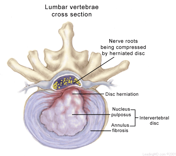
Patients with leg pain predominating (sciatica) may have
the diagnosis confirmed by an MRI of the lumbar spine or
a myelogram/CT scan. Two non-surgical treatments that can
be helpful:
- Epidural steroid injection
(ESI): The MRI may suggest that an injection of corticosteroid
(sometimes known as "cortisone") directly around the spinal
nerves, may be helpful. This is a special procedure. ESIs
are very safe, but the decision to have one should only
be made after a discussion with the physician.
- A selective nerve root block
(SNRB) is an injection which treats only one nerve. Frequently,
epidurals and selective nerve root blocks are done with
X-ray control to make sure the medicine is placed exactly
where it is needed.
Herniated Disc Patients With
Back Pain Predominating:
There are three main treatments for patients who have back
pain rather than sciatica:
- Exercise:The
mainstay of treatment for back pain is a good self directed
home exercise program to increase abdominal strength, back
muscle strength, and flexibility. There are many theories
on which exercises are best. A physical therapist trained
in back care will develop a personalized program with the
patient over a one to three week period. Good abdominal
strength is the key to a healthy back, therefore it is important
for the patient to continue these exercises indefinitely.
- Anti-inflammatory medicines
(NSAIDs):These are
often called arthritis medicines. It is important for the
patient to give them a full three to four week trial since
it takes this length of time for them to become fully effective.
There are many types, and each individual can probably find
one or two that work well.
- Bracing: If symptoms
persist over a long period of time, and exercise and NSAIDs
have not improved the condition, a brace may be worn to
provide additional support to the painful disc. When used
with a good abdominal strengthening program, a brace may
allow some people to be more active with less pain. Patients
should choose a brace that is comfortable enough to wear
for several hours at a time for the more strenuous activities.
Operative Treatment for Patients
with Leg Pain Predominating
The 20% who do not respond to treatment
after at least four to six weeks of non-surgical treatment and
a few who have special problems may benefit from surgery, such
as a microdiscectomy: a microscopic removal of
the disc rupture to decompress the pinched nerve. 
The indications for surgery include:
- Intense leg pain.
- The MRI shows a ruptured
disc compressing a nerve which is consistent with the distribution
of the leg pain.
- Testing the nerve by stretching
it ("nerve root tension signal") reproduces the
leg pain.
- There are no factors that
would make surgery a risk for the patient.
- Progressive worsening of nerve
function, such as any loss of bowel or bladder function.
The Surgical Procedure: Microdiscectomy
The operation usually lasts one to two hours and provides good
or excellent results in 95% of cases. Leg pain does not disappear
immediately after surgery, but gradually disappears over several
weeks.
- A general anesthetic is used,
and once asleep,the patient is placed in the prone or kneeling
position on a specially padded frame.
- A small incision is made
directly over the disc, and a microscope is then used to
find the compressed nerve and move it aside so that the
ruptured portion of the disc can be seen and removed. Only
the ruptured portion and loose pieces within about � inch
of the hole are removed.
- The space around the nerve
is then thoroughly examined to make sure no small pieces
of disc material might still compress the nerve.
- Finally, antibiotic solutions
are washed through the disc and incision to reduce the chances
of infection. An absorbable suture is used to close the
incision so that there are no stitches to be removed later.
What types of complications may occur?
All surgeries have risks, but complications
with this procedure are few. Nevertheless, it is important for
the patient to have a thorough discussion of these and other
potential risks with the doctor before making a decision to
have surgery.
- Scar tissue formation
(a 5% chance):When
this surgery fails, it is due to an overgrowth of scar tissue
around the nerve. Most people form some scar tissue in the
area of a surgery, but for unknown reasons, some individuals
form an extraordinary amount of scar which surrounds and
irritates the nerve. It can form along the spinal nerve
inside the spinal canal, or where the nerve exits the spine.
- Infection (a 3-5% chance):
Wound infection can happen any time an incision is made.
The bacteria can come from the skin around the incision,
the air in the operating room, or the bacteria that circulate
in the bloodstream. The steps taken in the operating room
to avoid infection are many. A dose of intravenous antibiotics
right before surgery reduces the risk of infection even
more. Persistent drainage from the wound 4-7 days after
surgery usually means an infection is present. The patient
might also have fever or chills, but this is not a reliable
indication of an infection. Antibiotics are usually successful,
but sometimes it is necessary to return to the operating
room to wash out the incision. An infection does not
usually cause the operation to fail, but may slow the healing
process.
- Spinal fuid leak (approximately
1%): Spinal fluid bathes the spinal cord and is contained
inside a sac called the dura mater. Sometimes
scar tissue forms between this sac and the ruptured disc.
A hole can develop in the dura when the surgeon is looking
for or removing the ruptured disc, allowing spinal fluid
to leak out. When a spinal fluid leak is encountered, the
hole is immediately repaired. Sometimes artificial blood
clot is added to form a seal around the repair. Usually
the patient is kept flat for 24 hours to allow the hole
to heal before resuming a normal recovery.
- Nerve damage at surgery
(<1% chance): A nerve that has been compressed by a
ruptured disc can be very fragile. Just moving the nerve
to get at the disc behind it might cause this fragile nerve
to be damaged. Fortunately, this is very rare, as is other
surgical damage to the nerve.
- Bleeding (rare):
Blood loss is a rare complication . People
whose blood does not clot blood normally are at increased
risk. The large blood vessels in front of the spine may
be damaged while removing disc material from within the
disc. This is extremely rare (perhaps one in ten thousand
cases) and requires emergency abdominal surgery to repair
the bleeding vessel.
- Recurrence (7%):
Though technically not a complication of surgery, there
is about a seven percent chance that the same disc will
rupture again, most likely in the first six weeks after
surgery when the hole in the disc annulus is healing. Even
after six weeks the disc continues to be prone to injury.
This is why the maximum weight a patient should lift
is 8-10 pounds for six weeks after surgery. Abdominal
strengthening exercises are recommended for life, since
strong stomach muscles are good insurance against recurring
disc problems.


© 2016 by LeadingMD.com All rights reserved.
Disclaimer