Shoulder Instability - Traumatic >> Diagnosis
How is a dislocation and traumatic shoulder instability diagnosed? As a rule, a sudden dislocation is quite evident. The patient usually
holds the arm against the side, since any attempts at motion cause
pain. A large crease under the acromion and a bulge in the armpit
are clues to the direction of the dislocation. However, when the
shoulder spontaneously relocates into its proper position, the diagnosis
can be more difficult. Patients may only report the feeling of having
the shoulder "slip" before the spontaneous reduction occurred.
A qualified individual usually can relocate the humerus at the site of the injury occurrence. Once the reduction is performed, there is immediate pain relief. Without medications, some patients may be unable to relax the shoulder muscles enough to allow the reduction to take place. Often, these patients must go to the emergency department to get the reduction accomplished.
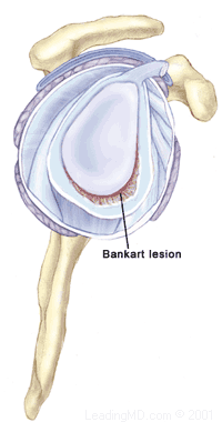
- X-rays are usually taken to confirm the dislocation, its direction, and to check for a related fracture. After the reduction, follow up X-rays will confirm proper positioning and assess any other injuries. X-rays may reveal a "bony Bankart", which is a fracture of the anterior-inferior glenoid (front, lower portion of the glenoid). The presence of this fracture indicates that the labrum and ligaments in the front part of the shoulder are no longer attached to the glenoid.
- If X-rays do not reveal such a fracture, an MRI or arthrogram may be ordered. In this diagnostic test, the status of the labrum and ligaments can be assessed. A Bankart lesion (detachment of the anterior-inferior portion of the labrum from the glenoid) is the most common cause of recurrent instability after an injury.